Unconventional nailing for tibial reconstruction in adolescent with osteogenesis imperfecta – A rare surgical case report
Ravikumar AS1*, Pradeep H2, Naveen HT2, Appu G Pillai3
1Professor, 2Assistant Professor, 3Postgraduate, Department of Orthopaedics
Sri Siddhartha Medical College, SSAHE, Tumkur,
Karnataka, India
* Corresponding author
Abstract
Osteogenesis Imperfecta (OI) is a rare connective tissue disorder known for excessive bone fragility caused by collagen mutations. The common orthopaedic problems related to bone fragility includefrequent multiple bone fractures, progressive deformity of long bones leading to impaired ambulation. Surgical correction of long bone deformities in OI is conventionally done using distraction osteogenesis (Illizarov fixator), elastic intramedullary nailing, rigid extramedullary fixation using plates, after osteotomy. Intramedullary fixation appears to be an ideal choice for correction of recurrent deformity in the long bone and the devices used previously include telescoping rods, single or dual non-elongating nails (rush nail, TENS). Recently reported use of humerus nails for femur reconstruction in 3 adolescent out patients. We report a case of humerus nailing for reconstruction of tibia in an adolescent OI male with excellent outcomes which is first of its kind and not reported previously by any other authors.
Keywords: Osteogenisis imperfect, Pathological fractures, Intramedullary nail, Deformity correction
Introduction
Osteogenesis imperfecta (OI) is a rare hereditary connective tissue disorder with the common clinical presentation of excessive bone fragility caused by mutations in collagen.[1] Clinical manifestations of OI are wide spectrum and could vary from lethal forms in the perinatal period to the subtle forms, which could be hardly identified and comparable to a normal person. However, the common orthopaedic problems in the OI patients, that is related to the excessive fragility, are frequent fractures, progressive deformity of long bones and impaired ambulation. The treatment of OI requires a multidisciplinary approach including both medical treatment, such as using bisphosphonates therapy to increase bone mineral density and reduce long bone fractures rate, and surgical treatment for fracture fixation or deformity correction. Goals of surgical management of OI are to minimize the incidence of fracture, restore bone axis and avoid deformity. The recommended surgical implant in OI is a load-sharing intramedullary (IM) device, with the largest diameter as possible, due to the better biomechanical property in fragile bone over plate construct and avoid plate-related complications, such as bony resorption from stress shielding, implant failure, progressive deformity and subsequent fracture at the plate ends. However, the surgical fixation in OI, especially in adolescent OI patients with tibial/femoral fracture or deformity, are still problematic due to particular abnormal bony anatomy (such as short limb, non-anatomical alignment secondary to previous injury/surgery, and narrow and non-linear medullary canal with super physiologic bowing) resulting in implant selection difficulty which is suitable for medullary canal size and bone length. Although there is an advanced surgical system, like a telescopic rod, which is specifically designed for OI patients with many different sizes that allow fixation in all age groups, this implant isn't available everywhere, including our country. Moreover, the traditional implant standard pediatric IM devices such as Rush pin/TENS might not be appropriate in these adolescent OI patients due to its small size and inability to provide rotational stability. Recently, there has been a few studies which reported that the small IM interlocking nail, such as humeral nail, could be used in femoral fixation in normal adolescent patients due to some advantages. The humerus nail being the IM locking device with smaller diameter and shorter length than conventional tibial nail which is appropriate for small-sized adolescent tibial anatomy, should also be suitable for tibial fixation in adolescent OI patients. It is commonly used for humerus fractures but here we report the first case of humerus nail being used for tibial reconstruction in these patients.
Case Report
A 14 year old boy with the history of recurrent lower limb fractures with trivial trauma presented to Orthopedic department with tibial deformity. He had undergone surgery for bilateral femur fractures. Tibial deformity noted after plating surgery. [Figure 1 & 2]
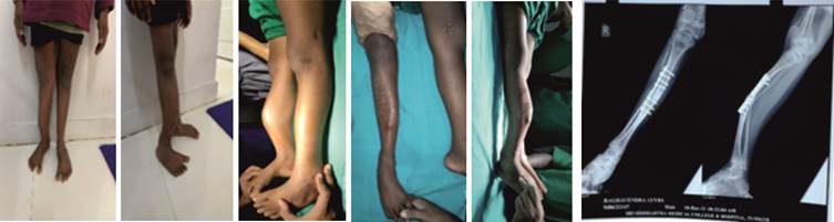
Figure 1(a-e) : Deformity in standing and supine position Figure 2: X-ray deformity
with plate in situ
Surgical technique
The patient was placed in supine position under spinal anesthesia and pneumatic tourniquet. Plate was removed through the previous surgery scar. Multi-level corrective osteotomy was performed with oscillating saw and osteotome. Medullary canal was recanalized with the help of drill bits of increasing diameter followed by hand reamers. Then the tibia was reduced to acceptable alignment. The entry point was made at the proximal tibia similar to the regular tibial nailing of adult bone, guide wire was passed in the Centro-medullary direction and the medullary canal was gently prepared by hand reaming using 6, 6.5 and 7-mm T-reamers. Then a 6.5-mm diameter Interlocking humeral nail (Sharma) with 240-mm length was inserted followed by proximal and distal locking screw insertion under fluoroscopic guidance. The bone gaps created due to the cortical splits during reaming were filled with bone grafts obtained from the anterior closing wedge osteotomy site. No additional wiring/implantation was performed. The operative time was two hours with minimal blood loss, and the total length of hospital stay was four days. [Figure 3]
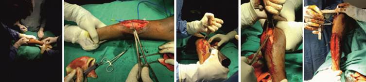
Figure 3(a): Incision (b) Osteotomy cut at diaphysis (c) Initial reaming with large drill bit
(d) Sequential rigid reamer (e) Nail introduction over corrected deformity
Postoperative care and rehabilitation
Sutures removed on 15th post operative day. Patient was allowed for knee and ankle ROM exercises and wheel chair mobilization for the first 6 weeks followed by weight bearing as tolerated. The proximal and distal osteotomy sites both showed clinical and radiological union in progress at six weeks follow up.
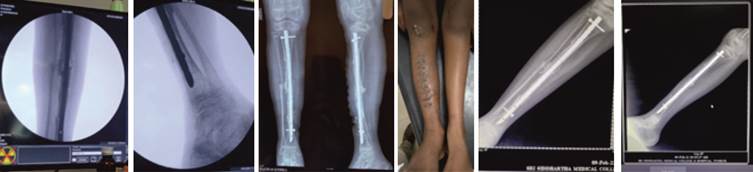
Figure 4 (a & b) : Intraoperative (c) : Final image after nailing (d) : Postoperative deformity correction
(e) : 6 weeks post-operative follow up
Discussion
OI is heritable disorder of collagen synthesis that commonly presents as bone fragility with multiple oslongum fractures and deformities. These bony problems usually require surgical management for fracture fixation or reconstruction by the load-sharing IM device.[6] However, the surgical fixation in OI patients, especially in the older children with tibial fracture or deformity, is very difficult due to the abnormal tibial anatomy resulting in implant selection problems such as a mismatch with conventional tibial IM nail, the poor fixation stability of standard pediatric IM devices, and the high cost of advanced telescopic rod. This study aimed to present the usefulness of humeral nail fixation as a surgical tool for tibial reconstruction in adolescent OI patients.
The humeral nail application for tibial fixation in adolescent OI patients has many advantages. Firstly, humeral nail, which is available in smaller diameter and shorter length than conventional tibial IM nail, is more suitable with these patient'stibial anatomy with narrow medullary canal and short limb. Secondly, humeral nail geometry has narrow width, it has a lateral bend at the proximal end which allows for insertion with minimal physeal violation, and 90-100 degree of the cephalomedullary angle for proximal locking blade/ screw. Thirdly, because of the interlocking nail property, the humeral nail would offer superior biomechanical benefits than gold standard Rush pin fixation as better in rotational stability and leg length control, especially for the patients with multilevel corrective osteotomy.
However, there were also some limitations for using humeral nail for tibial reconstruction in adolescent OI patients. Those are, however minimal, proximal tibialphyseal damage can not be avoided which predisposes for future growth disturbances of proximal tibia. Absence of Herzog bend limits the nail entry to a more proximal and posterior area of tibial articular surface.
The results of this study showed that using humeral nail implant in tibial fixation was possible and could be used in proximal, middle or distal location. Moreover, this feature can be indicated for fixation of fracture and nonunion, or corrective osteotomy. Our study also demonstrated the favorable outcome with 100% fracture healing and without the implant-related complications such as infection, nonunion, or Avascular necrosis. Therefore, we concluded that the humeral nail application for tibial fixation in adolescent OI patients is one of the possible options with satisfactory outcomes
Acknowledgement: NIL
Financial support and sponsorship: NIL
Conflict of interest: NIL
References
1. Burnei G, Vlad C, Georgescu I, Gavriliu TS, Dan D. Osteogenesis imperfecta: diagnosis and treatment. JAAOS-J Am Acad Orthop Surg. 2008;16(6):356–66.
2. Roberts TT, Cepela DJ, Uhl RL, Lozman J. Orthopaedic considerations for the adult with osteogenesis imperfecta. JAAOS-J Am Acad Orthop Surg. 2016;24(5):298–308.
3. Seikaly MG, Kopanati S, Salhab N, Waber P, Patterson D, Browne R, et al. Impact of alendronate on quality of life in children with osteogenesis imperfecta. J Pediatr Orthop. 2005; 25(6): 786-91.
4. Sa-Ngasoongsong P, Saisongcroh T, Angsanuntsukh C, Woratanarat P, Mulpruek P. Using humeral nail for surgical reconstruction of femur in adolescents with osteogenesis imperfecta. World J Orthop. 2017; 8(9): 735.
5. Esposito P, Plotkin H. Surgical treatment of osteogenesis imperfecta: current concepts. Curr Opin Pediatr. 2008; 20(1): 52–7.
6. Gil JA, DeFroda SF, Sindhu K, Cruz Jr AI, Daniels AH. Challenges of fracture management for adults with osteogenesis imperfecta. Orthopedics. 2017; 40(1): e17-22.
Address for Correspondence:
Dr. Ravikumar A.S.
Department of Orthopaedics, Sri Siddhartha Medical College, SSAHE Tumkur Karnataka India.
Email: dr.ravikumaras@gmail.com

Attribution-NonCommercial-ShareAlike
CC BY-NC-SA

An official peer reviewed publication of
Sri Siddhartha Medical College & Research Centre
Constituent College of Sri Siddhartha Academy of Higher Education
(Deemed to be University u/s 3 of UGC Act, 1956)
Accredited 'A' Grade by NAAC
Tumakuru, Karnataka, India. 572107
Research Journal of Medical and Allied Health Sciences is a medium for the advancement of scientific knowledge in all the branches of Medicine and Allied Sciences and publication of scientific research in these fields. The scope of the journal covers basic medical sciences, medicine and allied specialities, surgery and allied specialities, dentistry, nursing, pharmacy, biotechnology, public health and other branches of the allied health sciences. This journal is indexed with Advanced Science Index(ASI), National Science Library and Open J Gate.
E-ISSN : 2582-080X | : editor@ssmctumkur.org , info@ssmctumkur.org
Attribution-NonCommercial-ShareAlike 4.0 International (CC-BY-NC-SA 4.0)